We’ve Migrated!

Hello everyone,

Our apologies for the long break, we were working on a new project – We are happy to announce that we have expanded and merged our blog with a new website. This new site not only contains science posts from our lab, but every biology lab within the School of Life Sciences at UNLV. Most of our current posts have been migrated, and all new posts will appear on our new website instead of here. This blog will remain here for now, however, should we continue to post in the future.

Update your bookmarks and head on over to ScienceofSOLs.Wordpress.com

Thanks for reading!

List of Posts by Topic

Click any of the below titles to view that post, or scroll down to see posts in chronological order. Or, click on an author to the right to see that person’s posts.

Update

Graduate School

Science & Life in the Lab

Neurobiology

Articles Around the Web

Las Vegas Science Cafe

Misc.

The Graduate School Process

Author for this post: Travis Parsons, Ph.D Candidate (Apologies for the long hiatus, I had to take my comprehensive exam! What is that? Read on…)

In order to better understand science and trust in the scientific findings of researchers, it is important to understand the process by which these researchers become experts in their field. In this post, I want to spend a little time talking about the specifics of graduate school in the field of life sciences and what it takes to get a Ph.D. in biology. Hopefully this gives a better idea as to the process that scientists you see on T.V. or in the news go through in order to obtain their degree.

The first thing to know is that the process I’m about to describe varies in subtle ways from school to school, but the the overall path is quite similar. First, anyone wishing to pursue a Master’s or Ph.D. in Biology must first take an exam known as the Graduate Record Examination (GRE) (much like many other exams, the MCAT, SAT, ACT, etc.). Many schools also like to see prospective students take the biology subject exams that the same company offers. In biology, these scores serve mostly as a minimum threshold to surpass in order to be considered for admission into a graduate program. A much more important aspect of the application process is previous laboratory experience, college grades, and why one desires to pursue a higher degree in Biology.

During the application process, applicants are usually asked to identify professors that they are interested in working under. In biology, this is a very important task, as it potentially selects the lab that you will work in for the entirety of your graduate career. This professor will be your mentor, advisor, and boss for many years to come. The research area the professor specializes in will also become your future area of specialty. For this reason, many schools have 1 year rotations where new students work in several different labs in order to find the best fit for both the student and the professor. Almost all Ph.D. programs fly their prospective students out to the campus for free in order to interview both for admission to the program and for a proper fit within a lab (Master’s programs that do this are more rare – generally phone interviews are more popular).

Once a student accepts entry into a graduate program and selects their lab/advisor, their graduate career begins. Usually a student will take 2 years of classes while spending any other free time in the laboratory working on their research project. A student’s research project can either be determined by the student or the advisor (though in either case, by nature the student always takes it in their own direction by the end of the program). During these first few years, students must pass a rigorous set of exams that prove they are fit for the Ph.D program (Master’s programs generally don’t have these). These are known as Qualifying or Comprehensive Exams. They can vary in their timing – some schools give them in a student’s first year while others can be as late as the second or third. For example, students in the UNLV biology department’s “cell and molecular biology” group must take a 1-month exam in the summer of their second year. Students are given an original research paper that is outside of their area of knowledge and are asked to write an 11 page grant application that would further the research presented in the paper (as if you were the professor of that laboratory and were asked to get funding for whatever you would do next). Students have four weeks to become experts in this new field and must present two original experiments that have never been done before in real life. At the conclusion of the month, students must defend their proposals for 2-3 hours alone in front of a panel of 5 professors. (If you are a prospective student, don’t be afraid – You will be ready thanks to the training you have received over the past 1-3 years! This is what graduate school prepares you for). The point of these exams is not to torment students but rather to ensure they have the skills they will need to start their own careers in science and be able to get funding for their original research ideas.

Once a Ph.D. student completes their first 2 years of classes and passes their exams, they “advance to candidacy”. Usually this entitles them to a small pay raise and a new title of “Ph.D. Candidate”. From here, biology Ph.D. candidates will spend an additional 4 – 6 years researching their project and performing experiments in the lab or field until they are ready to graduate. While some graduate students are able to graduate in 4 years total time, the average is closer to 5 or 6 years. In order to graduate, students generally must publish their findings, write a thesis (if Master’s) or dissertation (if Ph.D.), and defend it publicly. For biology graduate students, the writing portion is generally less intimidating than it is for other fields, since you have plenty to write about after 4-8 years of laboratory experimentation.

After graduate school, many Ph.D.’s choose to do an additional 2-3 year period of “post-doctoral research” (where they become colloquially known as “Post-Docs”) where they usually join another lab/institution and work full time to try and generate more data and more publications in order to bolster their career prospects in the biotechnology industry or in academia. At this point, many biology Ph.D.’s choose to either go into either of these two fields, though many other routes exist, such as patent law or consulting. In a future blog post, I’ll bring up some interesting opinions that some big names in biology are sharing about how the structure of post-doctoral positions and labs in general needs to change in order to make the U.S. a better research powerhouse.

Hopefully this brief summary explains the long process by which researchers earn their Ph.D or Master’s degrees, and allows for a better view behind the training these scientists must receive before gaining these titles. Let me know if you have any questions about graduate school in the comments below, or if your graduate experience has been different! Thanks for reading.

Brainbows – Exploring Biology with Fluorescence

Author for this post: Travis Parsons, Ph.D Candidate

One immensely powerful tool in cell biology is that of fluorescence. Biologists have developed the ability to make cells glow almost any color under the rainbow in many specific ways. Want to make stem cells within the blood marrow glow green? What about have the nuclei of cells glow blue? Easily done in many model organisms! The ability to isolate cells by artificially ‘”coloring” them according to their identity or some aspect of their function or morphology has truly revolutionized biology. In the 1800’s, a man named Santiago Ramón y Cajal realized that he could use a special technique called Golgi’s silver nitrate staining to selectively label neurons at random within a brain. The human brain is a massively complex tissue that consist of ~86,000,000,000 neurons, resulting in a total of roughly 1014–1015 connections. There is no way simple observation could ever be able to tease apart the various computer-like networks and circuits that these neurons create. Santiago Ramón y Cajal made the first breakthrough into this realm by using this special staining procedure that labeled neurons at random. Now, only subsets of neurons could be observed rather than the whole neural network all at once.

A Golgi Stained Pyramidal Cell Source: http://www.wikidoc.org/index.php/File:GolgiStainedPyramidalCell.jpg
A Golgi Stained Pyramidal Cell. Note how only one neuron is stained within the context of an entire brain slice.
Source: http://www.wikidoc.org/index.php/File:GolgiStainedPyramidalCell.jpg

For the first time, Santiago was able to accurately draw the morphology of neurons.

A drawing by Santiago. Source: http://www.wikidoc.org/index.php/File:Purkinje_cell_by_Cajal.png
A drawing done by Santiago of a single Purkinje neuron. Note the many branching dendrites.
Source: http://www.wikidoc.org/index.php/File:Purkinje_cell_by_Cajal.png

Today, this technique has been taken further and further by researchers interested in mapping the entire “connectome” of the human brain (a map of the human brain’s neural connections). Unfortunately, Golgi staining only works so well: Labelling small subsets of neurons can never suffice to map out the entire brain. How can you tell that one neuron connects to another when you have to keep looking at different brain slices, from different animals, all of which have different neurons labeled, out of a possible 86,000,000,000? This is where recent advances in fluorescence and genetic manipulation come in. In 2007, Lichtman et al. dreamt up a way to label neurons with hundreds of different fluorescent colors. They named their ingenious technique “Brainbow”. Here’s how it works: Researchers have the ability to introduce foreign DNA into organisms, and DNA makes proteins. DNA can be engineered by scientists to contain instructions for making whatever protein they want. Several proteins have been discovered and engineered to fluoresce when excited by certain wavelengths of light. This means scientists can engineer an organism to make proteins that will glow different colors under a microscope equipped with the right light source. Today, all kinds of color options are now possible: Red, green, cherry, blue, tomato, ruby, yellow, you name it.

Lichtman et al. took advantage of this to engineer a piece of DNA that they could inject into animal brain cells to fluoresce different colors, thus labeling neurons distinctly so that they could be traced well enough to map their connections. The trick behind doing this is to inject DNA containing instructions coding for four different fluorescent proteins: Orange, red, green, and blue. At the same time, they used another special trick to make sure that only one of these color gets made per piece of engineered DNA that gets placed within a neuron. They did this with the help of a protein called Cre recombinase, which is capable of modifying DNA at specific sites within the genome. Scientists discovered this protein in a bacteriophage and then engineered it to be directly controlled and usable in other organisms that normally do not have it. In response to a special drug, this engineered protein turns on and can modify DNA at controllable locations. The researchers engineered their colorful DNA strand to be a target of this protein. What this means is that when the researchers apply a special drug to the neurons, Cre recombinase turns on and removes three of the four colors at random in each piece of engineered DNA within a neuron. This results in each neuron expressing only a single fluorescent color. What made this technique even better, however, is that the researchers realized that they could place several pieces of color-coding DNA inside each neuron. Now, each DNA piece can only express one color, but there are multiple DNA pieces in the neuron, and each ends up expressing one color at random. So one neuron can end up glowing red and blue at the same time. Or red and orange. Or red, orange, and green. The end result are neurons that express a whole spectral hue of colors that resembles a rainbow. Now, each neuron can be differentiated from it’s neighbor, and mapping can be achieved!


Example of a brainbow. Each neuron is a different colored fluorescent hue. Image sourced from Wikipedia at http://en.wikipedia.org/wiki/Brainbow. Original figure from Lichtman and Sanes, 2008
Example of a brainbow. Each neuron is a different colored fluorescent hue.
Image sourced from Wikipedia at http://en.wikipedia.org/wiki/Brainbow.
Original figure from Lichtman and Sanes, 2008
Example of a brainbow. Each neuron is a different colored fluorescent hue. Image sourced from Wikipedia at http://en.wikipedia.org/wiki/Brainbow. Original figure from Smith, 2007
Example of a brainbow. Each neuron is a different colored fluorescent hue.
Image sourced from Wikipedia at http://en.wikipedia.org/wiki/Brainbow.
Original figure from Smith, 2007

This gives you a small view into how fluorescence not only makes biology beautiful, but also is a critical tool in answering biological questions. We will have more posts soon on more ways in which researchers can directly manipulate genetics to study questions of human health and biology in general.

The importance of stupidity in scientific research

Author for this post: Travis Parsons, Ph.D Candidate

Here is an interesting article worth reading whether you are currently in graduate school, thinking about starting, or wondering why getting a Ph.D. is a different from other endeavors.


The importance of stupidity in scientific research by Martin A. Schwartz

Department of Microbiology, UVA Health System, University of Virginia, Charlottesville, VA 22908, USA


Why do research on model organisms?

Author for this post: Travis Parsons, Ph.D Candidate


Rather than perform experiments on humans, most research is focused on using other organisms as models for human biology. These organisms include the following:

Drosophila melanogaster (fruit flies)

Xenopus laevis (African clawed frog)

Danio Rerio (zebrafish)

Saccharomyces cerevisiae (yeast)

Caenorhabditis elegans (nematode)

Mus musculus (mouse)

Arabidopsis thaliana (Arabidopsis)

… and many more note listed here. (Link to Wikipedia list). If you would like to know more about why we use model organisms in general, see our earlier post here.


So just how good are these model organisms for approximating human health? The truth is that these models are extremely good models for answering questions about human biology, and about biology in general. The reason why is that all organisms share ancestry with one another through the process known as evolution. Many people are often confused about evolution’s basic premise. Evolution does not attempt to explain how life on Earth started, it only seeks to explain how life has evolved since it began. Scientists still do not know how life began, and evolution stands independently of this unsolved mystery.

On one level, this means that all organisms on Earth are directly related. On another level, this means that their molecular biology is also very similar (or “conserved”). This is a key concept to grasp when answering the question of how adequate these models are for human health. You may think you are pretty different from a mouse, but do you know how similar your liver cells are to a mouse’s liver cells? Or how similar a fruit fly’s insulin is to your insulin? How different would you believe your insulin is if we were to tell you that humans respond perfectly well to a fruit fly’s insulin and a fruit fly responds equivalently to human insulin? The two are interchangeable! Despite the myriads of differences between species, the cells that comprise these organisms are extremely similar. They attach to each other in similar ways, they talk to each other with similar signals, and they carry out the same basic cellular activities. Any modern FDA approved medicine you have ever taken in your life was given to model organisms first before human testing. Thanks to the evolutionary conservation between these organisms and humans, we can use them (responsibly, with proper research ethics and humane protocols in place) to create life saving pharmaceuticals, understand how different aspects of biology lead to disease, and how life as we know it works.

Unfortunately, more and more governmental science funding is going towards human-centric research and every year model organism researchers are receiving lesser and lesser funding. Despite the recent advances in human research, many human-only studies are less powerful than studies done on model organisms. Humans are complex, lead long lives, are subject to many environmental factors that affect their biology, and are impossible to control properly in studies (Example: It’s easy to give mice a fixed diet and see what happens… it is impossible to give human test subjects a strict diet and have them follow it for 5+ years). One recent technique has been to take two vastly different humans (for example, some one who is obese and some one who is extremely thin) and look for what genetic differences exist between them. It has turned out that genes are more variable than previously appreciated, and there are hundreds of differences that could explain these differences in biology. The only way to test which ones matter is to explore them in model organisms. It is important to note, however, that this a two way street. Because biology is so variable, there is also no way model organisms can ever replace human research. All findings in model organisms must be tested in humans or human cell lines in order to be applied as human medicine. But the truth is that the basic science is much more powerfully and convincingly done in model organisms before the successes are carried further to human trials. Hopefully this post begins to explain why both basic science research and model organism research are critical to improving human medicine and health – there is no faster or more powerful way to research biology and apply it towards humans. Our blog will continue to have further discussions on this topic as we get more in depth about these specific ideas. Please continue to support funding for basic scientific research and model organisms. Our findings will continue to pave the way for human medical scientists to bring these findings to clinical trials, and eventually to you and your family.


Surviving the First Year of Grad School

Author for this post: Sheila Mosallaei, Ph.D. Student.

The first year of grad school is similar to the feeling of being the new kid in middle school that comes in half-way through the school year. Everything is new and foreign to you, and yet everyone else knows exactly what is going on. Entering grad school is nothing like entering the first year of undergrad. During the first year of undergrad, there are hundreds of new incoming students fresh from high school eager to meet new people, learn new things and experience the infamous “college life”.   Grad school is a bit different. The new incoming class is much smaller, the students are all in different majors, and are all entering into different labs. In the beginning, it can be a bit intimidating. Lucky for me, I entered a lab very welcoming of me upon my arrival, making my transition smooth and steady.

During your first semester, it is difficult to accept that for the first time in life, classes are not your number one priority. Moving from the classroom setting into a lab is the biggest change when entering grad school. Your main focus becomes your research. You find yourself reading more and more papers, hoping that if you read enough, it will all magically start to make sense. Grad school is filled with opportunities to learn about everyone else’s research and learn about the new progressions in the science world. At first, going to seminars, colloquium, journal club, lab meetings and other new experiences will feel as if everyone is speaking another language and you are the only one who needs Google Translator. This feeling will go away (so I’ve been told).

Throughout this time I have been in grad school, I have come to learn the best way to progress is communication. By communicating more with my lab mates and asking them questions about everything, I have come so far in just a couple months. I have come to learn the best resource for information is no further than inside of your lab. Spending time in the lab is key to progress in grad school. Although I am only in my second semester of grad school, I have already learned so much. Even if at times you begin to feel distraught, discouraged, or about to lose your mind, it is important to remind yourself of the perks of being in grad school: they provide free food at the speaker events.

How are Drosophila reared in the lab?

Author for this post: Travis Parsons, Ph.D Candidate

One inconvenience of rearing Drosophila is that since they carry unique genetic mutations that often take large amounts of time and effort for researchers to generate, labs chose to maintain many of their flies (or “stocks”) indefinitely rather than let them die. This is especially true in the case that a certain fly strain has been published in a scientific paper. Labs have the responsibility of keeping that genetic line alive so other researchers may request it, or use it in their research, or characterize it further. This helps to prevent scientific fraud.

Keeping flies alive takes a significant amount of time and effort for researchers. In our lab, every senior lab member spends roughly 2 hours keeping their flies alive per week. This also carries the drawback of having to plan vacations and time off carefully – the flies don’t stop their development to wait for you to return.

Many labs in the past used to raise flies in glass bottles, but this is very inconvenient when you have thousands of stocks and have to wash every bottle. Now, labs use plastic vials and bottles that are disposable. Seen below is a typical vial we use in our lab with a quarter for scale. The top is plugged by cotton or rayon, which prevents flies from escaping the vial (as well as keeps other flies out – remember, every fly strain is a purebred mutant. If any other fly gets in, the entire stock is contaminated. We keep 2 copies of our stocks in case an issue arises with 1 copy).

20150120_084331

Even the plastic used for rearing flies is an important consideration. Other options exist that are more translucent, but these vials are more brittle and food dries out more rapidly in them.

20150120_084420

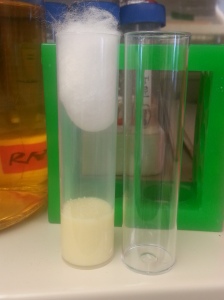
The bottom of the vial is filled with Drosophila food, which is what the flies and larvae eat as well as what they lay their eggs in. Our lab makes giant batches (Tens of liters) of fly food every week that consists of water, yeast, soy flour, cornmeal, agar (essentially a gelatin like substance), light corn syrup, and propionic acid. We also sprinkle extra yeast on top of the food.

20150120_085723

We keep large stocks of our fly food reagents on hand, as we go through them rapidly. We freeze them to prevent other organisms such as mites from contaminating our cultures.

20150120_085745

When we want to grow larger amounts of flies in one vessel, we use bottles.

20150120_085146

When flies are first ‘flipped’ or ‘pushed’ into a new vial, it looks something like this:

20150120_090608

From this point, the rate at which the flies develop depends on the temperature you keep them at. Most fly labs have sets of incubators at 18°C, 22°C, and 25°C. At 18, flies take ~18 days to develop to adulthood. At 22, it takes ~11 days. At 25, flies take ~9 days. This means most flies can stay in 18 for long term storage for about a month before we must change their food and vial.

During these time periods, the adult flies lay eggs in the food. These eggs hatch and larvae emerge. The larvae crawl into the food and eat large amounts of food as they exponentially increase in size. During this time, cells that will later become the adult fly begin to multiply. Afterwards, the larvae crawl out of the food and up the sides of the vial, where they turn into pupae and begin to metamorphose. Days later, adult flies emerge from the pupal cases.

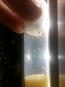
The below picture shows a vial that has these life stages present in it.

20150120_105623

The white objects on the walls of the vial or bottle are larvae that have crawled up the side of the vial and are ready to pupate. The darker objects are flies that have already done so. The lighter food at the bottom is food that no larvae have been able to reach or eat, while the darker food on top is full of larvae eating and churning up the food.

Over time, the food is used up and the vials become crowded and flies must be flipped into new vials. At this point, older flies have died, and younger flies have been born. They are transferred and the process begins anew.

This is multiplied across every fly stock a lab maintains, which are organized in racks.

20150120_092628

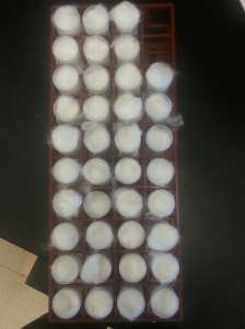
Some universities such as Indiana University have a Drosophila stock center where they maintain massive amounts of stocks for other labs to requisition. At the time of writing, the Bloomington Stock Center lists 58,967 stocks that are available for order. Assuming they have backups which they surely do, that doubles to 117,934 stocks. That’s 2,945 of the racks in the above picture. If they spend only 30 minutes flipping the stocks in each rack, that means the Bloomington stock center alone spends ~1,474 hours (or ~48 days) flipping their flies every month.

Hopefully this provides a window into how Drosophila are reared in the lab, and why it takes a significant amount of time, money, and effort.

What are you working on?

Author for this post: Travis Parsons, Ph.D Candidate

Over time, projects within a laboratory evolve as the questions a lab is interested in change. To get a nice perspective on what the Raftery lab has been researching for the past few years, check out our research page here.

This post will hopefully give a more generalized view of what we are interested in. During your life span, your cells are constantly communicating to one another and rearranging themselves. Many people tend to think of two things when they hear that cells can move/migrate. One is that this is clearly very important during development, where you transition from being a single cell into an organism with trillions of cells. The other is that when cancerous cells move (termed ‘metastasis’), it can be deadly. What most people forget to appreciate, however, is that cells move all the time, throughout your entire lifespan. Cells reorganize when you heal from a wound. Neurons move to remodel your brain as you learn information. During angiogenesis (the creation of new blood vessels), cells are moving to create new tissues. As a result, cellular movements are critical for development, normal maintenance of your body, and in disease.

The next idea to appreciate is that cells don’t just move on a whim. Think about how compartmentalized your body is. Your digestive system prevents any food or digested matter from going where it isn’t supposed to. Your circulatory system keeps blood where it is needed and not elsewhere in your body cavity. Your brain is protected by a blood-brain barrier that keeps unwanted infections and molecules out. Your skin keeps everything contained within you. This is achieved by cells that work together very coordinately. If even a couple cells failed to do so, the results would be disastrous: Internal bleeding, septic shock, infection, and more. For this reason, cells aren’t just inanimate objects. They communicate with one another all the time – sometimes hundreds of times per second. They send messages via chemicals, proteins, electrical currents, and possibly much more we haven’t discovered yet. These messages between cells tell them if they should move, when, how often, where to, when to start, when to stop, and much, much more.

So what is it that we are interested in? We wish to know how cells are communicating information about their movements to one another. If we can understand what signals tell cells to migrate, we can begin to understand many of the things we introduced above (how development proceeds, how to prevent cancer metastasis, how to enhance wound healing, and more). Many labs are looking to answer these questions by using human cell lines (cells that are derived from humans) but we are taking a different approach wherein we try to understand them in the context of entire tissues (for example, rather than study kidney cells by themselves, study the entire kidney as a tissue). To do this, we are using the ovary of the common fruit fly, Drosophila melanogaster (Why are we using a fruit fly? See our last post). We are able to dissect the ovary out of the fruit fly, and then culture it artificially for hours at a time. During this time it develops normally, despite being outside of the body of the fruit fly, and we can observe how the cells rearrange and reorient themselves within the context of an entire tissue. By using the awesome power of Drosophila genetics, we can proceed to genetically manipulate any part of their genome (in particular, genes that play a role in signaling cells to move) and observe the resulting defects that occur. By using this approach, we will be able to genetically dissect what genes are responsible for producing the signals that govern cellular movement. Over time, our lab as well as others will begin piecing together the signaling network that cells use to migrate, and how or why these signals go wrong during disease.

Hopefully this post provides a brief insight into what our lab is working on. More posts to follow soon!